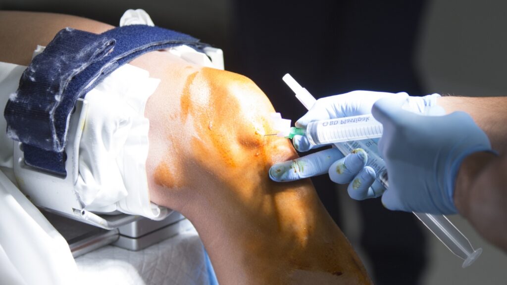
Knee arthroscopy is a minimally invasive surgical procedure used to diagnose and treat problems inside the knee joint. It involves using a small camera, called an “arthroscope,” to view the inside of the knee. The camera is inserted through small cuts in the skin, usually no bigger than a pencil eraser. This allows the doctor to see what’s going on inside the knee without making a large incision.
The patient is usually given anesthesia, which can be either local (numbing the area) or general (putting the patient to sleep), depending on the complexity of the procedure.
Making small incisions
The surgeon makes tiny cuts around the knee joint, through which the arthroscope and other small surgical instruments are inserte
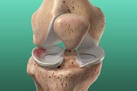
Viewing the knee
The arthroscope sends real-time images of the knee joint to a monitor, allowing the surgeon to examine the cartilage, ligaments, and bones. This helps identify issues like tears in the cartilage, ligaments, or inflammation.
Treatment
If the surgeon finds a problem, such as a torn ligament or damaged cartilage, they can use the instruments to repair or remove damaged tissue. These instruments are thin and specially designed to be used through the small incisions.
Recovery
Since the incisions are small, recovery time is typically quicker compared to traditional surgery. Most patients can go home the same day and are advised to rest and follow a rehabilitation plan to regain strength and movement in the knee.
Knee arthroscopy is commonly used for conditions like
Torn meniscus (cartilage)
The meniscus cushions the knee, and tears can cause pain and limited movement. Arthroscopy allows the surgeon to repair or trim the tear.
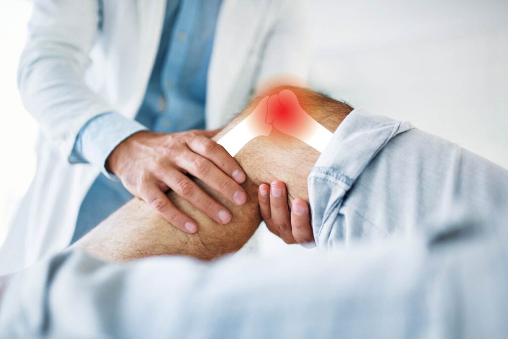
Ligament tears (like ACL injuries)
ACL tears, often from sports, can destabilize the knee. Arthroscopy helps repair or reconstruct the ligament, restoring stability.
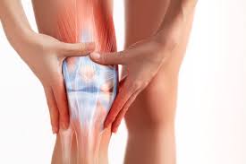
Knee joint inflammation or infection
Conditions like arthritis or infection can cause pain and swelling. Arthroscopy allows the surgeon to clean out infected or inflamed tissue.
Removing loose fragments of bone or cartilage
Fragments can cause pain and restrict movement. Arthroscopy removes these fragments for smoother joint function.