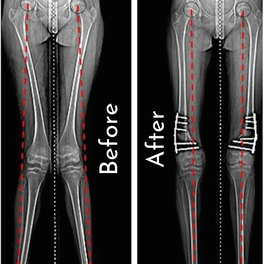
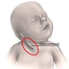
Torticollis (Congenital Muscular Torticollis)
Procedure: Treatment typically starts with physical therapy; if ineffective, surgical intervention may involve lengthening or releasing the sternocleidomastoid muscle.
Explanation: Torticollis causes the head to tilt to one side due to tightness in the neck muscles. Surgery, when necessary, helps restore normal head position and prevent permanent neck deformity.